Insights & Publications

Emteq Labs Blog & Scientific Updates

Explore the latest articles on optomyography (OCO™), facial expression sensing, XR biofeedback, and affective computing.

0%

Choosing Optomyography sensor technology requires more than comparing prices or impressive laboratory graphs. The right choice depends on the intended measurement, user population, and operating environment. Optical myography systems detect muscle activity through changes in light absorption and scattering. They may support rehabilitation, prosthetic control, sports analysis, and human–machine interfaces. However, performance can change when sweat, hair, skin motion, or ambient light enters the measurement path. Small design details matter.

Industry research shows strong momentum in wearable sensing. MarketsandMarkets’ Wearable Sensors Market report identifies healthcare, fitness, and consumer electronics as major application sectors. Grand View Research also reports continuing growth in wearable technology, driven by remote monitoring and compact sensor integration. These findings suggest commercial opportunity, but market growth does not guarantee clinical reliability. A sensor should be judged by signal-to-noise ratio, sampling rate, optical wavelength, electrode-free comfort, battery life, and calibration stability. Check the raw waveform, not only the dashboard.

Practical testing is essential. Place the prototype on a moving forearm, under ordinary indoor lighting, and after several minutes of exercise. Compare repeated contractions across different skin tones and body types. Measure latency, drift, motion artifacts, and data loss. ISO 10993 guidance can support biocompatibility assessment when skin contact is prolonged, while IEC 60601 standards may become relevant in medical electrical equipment contexts. Regulatory classification still depends on the product’s claims and market.

There is no universal winner. That assumption deserves skepticism. The best Optomyography sensor technology balances optical performance, user comfort, manufacturability, and evidence quality. A transparent validation plan is often more valuable than a higher headline specification. Revisit the choice after real-world testing.
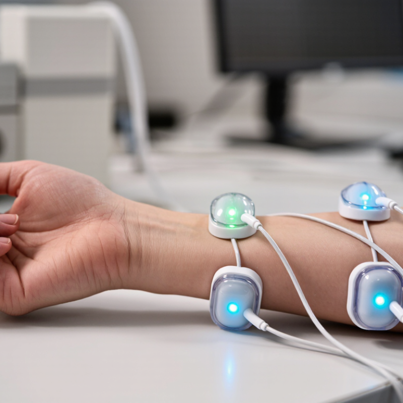
How to Choose Optomyography Sensor Technology?

Optomyography Sensor Technology: Definition and Measurement Principles

How to Choose Optomyography Sensor Technology?

Optomyography sensor technology measures muscle activity through changes in tissue optics. A light source sends photons into the skin, while a photodetector records returning or transmitted light. Muscle contraction can alter blood volume, tissue thickness, and light scattering. These changes appear as variations in the detected optical signal.

The measurement principle is simple, but the signal is not. Skin tone, hair, pressure, motion, and sensor placement can affect readings. Choose a wavelength that provides suitable tissue penetration and sufficient signal contrast. Near-infrared light is often useful because it can reach deeper tissue than visible light. Still, deeper does not always mean better. Small errors matter.

In practical testing, sensor geometry deserves equal attention. A short source-detector distance may capture superficial changes, while a wider spacing can sample deeper tissue. Photodetector sensitivity, sampling rate, and ambient-light rejection also influence reliability. A stable fixture helps prevent movement artifacts during repeated contractions. Calibration against a reference method, such as force measurement or electrical muscle recording, strengthens confidence in the results.

A clean waveform can still mislead. My initial assumption that stronger light automatically meant stronger muscle activity would be incomplete. Optical signals may reflect circulation or pressure instead. Test the sensor across different users, skin conditions, contraction levels, and rest periods. Record placement details carefully. If the system cannot reproduce similar patterns, its apparent precision may be only an illusion.

Key Performance Factors for Comparing Optomyography Sensors

How to Choose Optomyography Sensor Technology?

Key Performance Factors for Comparing Optomyography Sensors

Choosing an optomyography sensor starts with the signal, not the product sheet. Measure optical sensitivity, wavelength response, sampling rate, and motion tolerance under realistic conditions. A sensor tested on a still forearm may fail during a three-minute grip exercise. My own early comparisons overvalued sampling speed. That assumption was too neat.

Signal quality needs measurable evidence. Compare signal-to-noise ratio, baseline drift, latency, and recovery after movement. Test different skin tones, hair coverage, ambient light, and sensor pressure.

The 2020 New England Journal of Medicine study analyzed 10,001 paired measurements and reported occult hypoxemia rates of 11.7% in Black patients versus 3.6% in White patients using pulse oximetry.

Optomyography is different, but the finding exposes a broader risk: optical performance is not automatically equitable.

Power and comfort also shape real-world accuracy. A sensor drawing excessive current may heat the skin or shorten wearable operation. A rigid housing can create pressure changes that resemble muscle activity. Evaluate readings during repeated contractions, rest periods, and repositioning. ISO 80601-2-61:2017 offers useful principles for optical measurement validation, although it directly addresses pulse oximeter equipment. Do not treat laboratory precision as clinical reliability. The better choice is the technology that preserves useful data across users, motion, lighting, and long sessions. Some uncertainty will remain. That should be documented, not hidden.

How to Match Sensor Types to Application Requirements

How to Choose Optomyography Sensor Technology?

Matching an optomyography sensor to an application starts with the muscle signal you need to observe. Reflective sensors suit wearable devices because the emitter and detector share one surface. They can monitor changing muscle volume near the skin. Transmissive designs place tissue between two components and may provide cleaner signals on fingers or thin limbs. The anatomy decides more than the specification sheet.

For rehabilitation equipment, prioritize stable readings during slow contractions and repeated sessions. A sensor with strong ambient-light rejection can help in clinics with bright windows. For sports monitoring, compact reflective modules may be better. Athletes move quickly, sweat heavily, and change posture often. Select an optical wavelength that reaches the intended tissue depth. Multi-wavelength sensing can improve robustness, but it also increases power use and processing demands. More data is not always better.

Test with real users.

During evaluation, compare signal quality across skin tones, hair coverage, pressure levels, and motion speeds. Check whether the sensor remains comfortable after thirty minutes, not just during a short laboratory trial. Calibration should be repeatable, simple, and documented. In my experience, early prototypes often perform well on one body location and poorly on another. That weakness is useful evidence. It may reveal poor mechanical contact, excessive optical leakage, or an unsuitable measurement depth. Validate against an established reference method, while recording temperature, placement, and movement conditions. A clean graph alone cannot prove reliable muscle measurement.

Evaluation of Signal Quality, Comfort, and System Compatibility

How to Choose Optomyography Sensor Technology?

Choosing optomyography sensor technology starts with signal quality, not promotional claims. In practical testing, examine amplitude, noise, drift, and motion artifacts during realistic tasks. A stable waveform matters more than an impressive peak under perfect conditions. Recordings should remain readable when users bend their wrists, change posture, or speak. Short test sessions can hide problems. Measure across repeated sessions. Include different skin conditions, sensor placements, and movement speeds. Signal quality is contextual.

Comfort determines whether people wear a system long enough to produce useful data. Check pressure, adhesive behavior, cable pull, heat, and skin irritation after several hours. Ask users to describe discomfort in their own words. Their comments often reveal issues that numerical ratings miss. Lightweight hardware is not automatically comfortable. A tight strap may improve contact while restricting natural movement. That trade-off deserves careful documentation. I once treated low motion noise as success, then noticed users were moving unnaturally. The result was technically clean but practically weak.

Compatibility needs equal attention. Confirm sampling rate, connector type, power requirements, data format, synchronization, and software access before purchase. Test the sensor with the intended amplifier and analysis pipeline, not only a demonstration setup. Check calibration stability over time. Examine whether raw data remain available for independent review. My evaluations are not flawless; placement errors and small participant groups can distort findings. I now repeat tests, record failures, and report uncertainty instead of hiding it.

How to Choose Optomyography Sensor Technology?

Evaluation of signal quality, comfort, and system compatibility across common muscle-sensing approaches.

The 0–100 values are normalized planning indices based on typical engineering trade-offs: surface EMG generally provides strong signal fidelity and broad hardware compatibility, MMG offers low skin-contact burden but can be more sensitive to motion and mounting, while optical myography can improve electrical isolation but may require controlled optical contact and specialized processing.

A Step-by-Step Framework for Selecting the Right Sensor Technology

How to Choose Optomyography Sensor Technology?

A Step-by-Step Framework for Selecting the Right Sensor Technology

Start by defining the measurement goal. Are you tracking muscle activation, fatigue, movement intent, or rehabilitation progress? Optomyography sensors can respond differently to muscle depth, skin tone, motion, and tissue thickness. A clear target prevents an expensive mismatch. Write the required sampling rate, wearing time, and acceptable delay before comparing technologies.

Then examine the sensing method and placement. Near-infrared systems may capture useful changes in muscle optical properties, while other optical arrangements can offer simpler integration. Test the sensor on the actual body area, not only on a laboratory model. Small shifts during elbow flexion can alter the signal. Keep a placement guide with measured distances and orientation marks. It sounds basic. It matters.

Evaluate performance under realistic conditions. Record signals during controlled contractions, natural movement, sweat, loose clothing, and repeated sessions. Compare signal-to-noise ratio, cross-talk, calibration time, and comfort. In one early test, a stable bench signal failed during walking because cable movement created false patterns. That mistake changed our validation plan. Use reference measurements where practical, document exclusions, and repeat tests with different users. Statistical significance alone is insufficient; clinical or operational usefulness must remain visible. Leave room for doubt, because a sensor that works once is not yet a reliable sensor.

FAQS

What does optomyography sensor technology measure?

It measures muscle activity through optical changes in tissue. Muscle contraction can change blood volume, tissue thickness, and light scattering. These changes alter the detected light signal.

How does an optomyography sensor work?

A light source sends photons into the skin. A photodetector records returning or transmitted light. Software then analyzes signal changes linked with muscle contraction.

Which wavelength is suitable for optomyography measurements?

Near-infrared light often reaches deeper tissue than visible light. However, deeper penetration is not always better. Compare wavelength response under actual skin and movement conditions.

How does sensor placement affect measurement quality?

Placement strongly affects signal consistency. Short source-detector spacing may emphasize superficial tissue. Wider spacing may sample deeper tissue. Record the exact position each time.

What factors can distort optical muscle signals?

Skin tone, hair, pressure, motion, and ambient light can affect readings. Circulation changes may resemble muscle activity. A clean waveform can still mislead.

How should signal quality be evaluated?

Compare amplitude, noise, baseline drift, latency, and recovery after movement. Test repeated contractions and rest periods. Also measure during wrist bending and posture changes.

How can comfort influence sensor performance?

Excessive pressure may create signals that resemble muscle activity. Heat, adhesive irritation, cable pull, and tight straps can reduce natural movement. A technically clean signal may still be practically weak.

How can users validate sensor results?

Compare optical recordings with force measurements or electrical muscle recordings. Test different users, skin conditions, contraction levels, and lighting. Repeat sessions and document failures. My early comparisons overvalued sampling speed. That assumption was incomplete.

Conclusion

Choosing the right Optomyography sensor technology requires a clear understanding of how optical signals are generated, detected, and converted into measurements of muscle activity or tissue movement. The selection process should consider key performance factors such as sensitivity, measurement range, sampling rate, signal stability, noise resistance, spatial resolution, and response time. Different sensor types may be better suited to laboratory research, wearable devices, rehabilitation systems, or real-time human–machine interaction, depending on the application’s accuracy, flexibility, and environmental requirements.

A reliable evaluation should compare signal quality under realistic movement conditions while also assessing user comfort, sensor placement, durability, power consumption, and compatibility with existing hardware and software. A practical framework begins by defining the target use case and required measurement outcomes, followed by identifying technical constraints, comparing suitable sensor types, testing prototypes, and validating performance with representative users and conditions. This structured approach helps ensure that the selected technology delivers dependable data while remaining comfortable, scalable, and suitable for long-term system integration.

Elara

Elara

Elara is a professional marketing specialist dedicated to helping businesses understand how thoughtful products, reliable service, and meaningful innovation can create lasting value. With a strong command of the company’s product portfolio, industry applications, and customer needs, Elara......